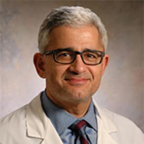
Dr. Husam Balkhy, Associate Professor of Surgery at the University of Chicago Medicine, discusses part one, of a two part lecture, on the minimally invasive and robotic heart surgery program at the University of Chicago Medicine.
[MUSIC PLAYING] HUSAM BALKHY: Good afternoon. My name is Sam Balkhy. I'm an associate professor of surgery and the director of the minimally invasive and robotic cardiac surgery program at University of Chicago Medicine. I wanted to give you a brief overview this afternoon on the minimally invasive and robotic heart surgery program at the University of Chicago Medicine and the variety of surgical procedures that we perform using these relatively innovative and new techniques. For starters, I want to boast about our new hospital, which opened in the spring of 2013-- the Center for Care and Discovery, which is an apt name for it because it discovers the innovation that we have and it is providing a lot of great care for a lot of our patients. The program for robotic and minimally invasive surgery began in July of 2013. And we have a great team and perform all sorts of different types of procedures using the da Vinci robot and other minimally invasive techniques for a wide range of patients from all over the country and all over the world really. I'll start off by talking a little bit about what it is that propels us to do minimally invasive heart surgery. The first thing is that the surgeons mandate is to minimize trauma. We've perfected a variety of techniques to fix the heart from some of the ailments that happened to it-- holes in the heart like ASDs and VSDs, mitral valve leaks, coronary blockages. We've basically perfected the techniques to fix those problems with a wide-open sternotomy. And that represents the efficacy and the gold standard of what we do. The rationale as surgeons today is to try to improve this procedure by reducing morbidity and reducing the time that it takes for a patient to heal, and the time that the patient is out of work and recovering and the body is somewhat compromised. And so we're trying to improve recovery time, minimize wound complications, improve on bleeding as well as cosmesis, and indeed the quality of life after these procedures. So why would we want to use robotics? Well, it turns out that most cardiac surgical procedures require a level of dexterity and precision that is more than what is allowed by traditional laparoscopic and thoracoscopic techniques. So the dexterity of the da Vinci robot is what is required for some of these very complicated procedures. There have been multiple studies to look at the quality of life after heart surgery in terms of whether it's done with a sternotomy or whether it's done with a sternal sparing approach. And many of these studies have shown an earlier return to work, an increase in the vitality at an early phase after surgery, improved mental health and social function very early on after heart procedures. This is a slide that shows that patients return to work after a mini thoracotomy sternal sparing incision for coronary bypass relatively quickly, within four to five weeks, compared to 10 to 15 weeks if they have an open procedure. The da Vinci robot is a well-known entity at University of Chicago Medicine. We perform multiple non-cardiac procedures with this device as well. It basically allows the surgeon to have a natural three-dimensional operative field with aligned instruments and manual dexterity that is very similar, indeed better, than what the human hand can provide. It is a very, very sophisticated piece of technology that has come a long way and has had multiple iterations and multiple generations of it become available. There have been significant regulatory milestones. We've been doing cardiac and thoracic surgery with this device for over 10 years now. It's surprising to some that this is a new technology. And they think that it's relatively new and only for a couple of years. But indeed the FDA approved the da Vinci robot for cardiac surgery back in 2002, so about 12 years ago. And many, many surgical procedures have been done with it. Here's an example of what the device can allow us to do. These are microscopic instruments. And you can see that the robotic arms allow for three instruments in this case. So it turns a surgeon's hand from having two hands into having the ability to have three hands. And so the toothpick is being held by one hand. And then, there's a very fine scissors and a very fine microscopic forcep that allows the grape, as it were, to be peeled. And you can see how the surgeon manipulates these instruments inside the patient's body from a remote console, reproducing the actual movements at the console and then having the instruments repeat those. Obviously, the robot is a passive participant in this. It does not do the procedure. The surgeon does the procedure. That's one of the biggest fallacies. Sometimes, a patient thinks that a small robot goes into their body and does the operation. That's not the case. This is an example of a comparison between a patient who had a sternotomy incision and another patient who had a smaller incision for the same operation. And you can see that the difference can be dramatic, if there's a keloid, for example. So what are the procedures that we do with the da Vinci robot at University of Chicago? Well, I try to divide them into three main categories. The first category is intracardiac, meaning procedures that we do inside the heart that require us to put the patient on the heart/lung machine, require us to stop the heart in some or the majority of cases. And these really kind of fall into surgery on the mitral valve, in addition to the tricuspid valve. So we do mitral valve repair or double valve surgery-- mitral and tricuspid-- and then repair of septal defects, removal of myxomas, and removal of other masses potentially from the heart. This is a diagram just to show that the mitral valve is best accessed from the right side of the patient. Today, most heart surgery is done through a sternotomy. And that's just by default, because most surgeons learned how to operate on the heart doing coronary bypass. And it's best to do coronary bypass through the front, because you can access all the different areas. But mitral surgery began a long time ago, and surgeons were operating from the right chest. We've learned now to minimize the incisions, but also maintain the access from the right side and are able now with instrumentation and endoscopic techniques to see the valve in its entirety-- both the super- and the sub-valvular apparatus-- all from the right side, which is the way the valve is oriented. So that allows us, in general, to perform repairs more often than replacements, which is really what the gold standard is. This is what the setup looks like in the operating room for a mitral valve repair. We have a two sonometer working port in the fourth interspace and multiple other small 8-millimeter to 12-millimeter ports that contain the robotic arms and the robotic camera. The cannulation is in the groin. And you can see here on this video, a very, very small groin incision. This is the patient's right groin. And basically what this is showing is the very fine dissection of the femoral vein and the femoral artery to allow us to place the patient on the heart/lung machine. And these now are the ports that we're using to position the robotic camera and the robotic instruments. And you can see how small these little incisions are. And this is really all that the patient is left with. There are no big incisions. There's no rib spreading. The morbidity from this is very, very minimal after the surgery is done. The working port, as it were, which is what this is called, allows the assistant at the table to administer the instrumentation and the rings and the sutures that are required for the procedure. Once the patient is setup for this operation, this is some video of the mitral valve being seen and examined with the robotic instruments and the endoscopic apparatus. And this provides a surgeon with a three-dimensional 15 times magnification view of the mitral valve. And this patient happens to have a torn cord in the posterior leaflet of the mitral valve and is undergoing a mitral valve repair. And the way that we repair valves these days is mostly to replace the torn or the elongated cords and do an annuloplasty to the ring. And these cords can be replaced through the anterior leaflet or to the posterior leaflet. Here, you can see the cord being attached to the papillary muscle. We get a great view of the papillary muscle, which is inside the ventricle and is sometimes very difficult to see from a sternotomy approach. And then, the cord gets attached to the edge of the leaflet that is prolapsed. And then, using a very accurate technique of filling the ventricle, we adjust to the length of the cord. Here, as you see, we fill the ventricle with water, and we're tightening up the cord to measure exactly how long it needs to be. And then, once we seal the leak as you see here, then we know that it's time to tie the cord and leave it at that length. And you can see in the last picture there, the torn cord is still attached. We haven't cut it off yet. And so that basically allows us to with a lot of precision replace cords and measure them perfectly well. And this is what the final outcome will look like to the bottom on the right there. We have a ring that is applied to size down the annulus and allow the two leaflets to come up against each other and prevent the leak. So this is a perfect example of a straightforward mitral valve repair. But this can be done to more than one torn cord. It can be done to the tricuspid valve as well. It can be done by resecting some areas of the leaflet that are elongated or torn. This is an example here of a patient who's had previous mitral valve surgery. And she had a mitral valve repair in the past and developed a recurrence of a mitral valve leak. And with using the da Vinci robot, we're basically repairing her posterior leaflet in a different way. We're augmenting it, and we're using a patch to basically make the posterior leaflet a little bit more voluminous to be able to become competent. And this is a patch made out of extracellular matrix. And you can see the robotic instruments allow us to really get in there and sew this patch on and make the valve competent again. So just a look at what the varieties of the procedures that we can do. Redoes are a little bit harder, but definitely doable, especially in the mitral valve surgery as well as tricuspid valve. Sometimes, there are adhesions around the lung that we can easily dissect off and come back and get access to the valve as you see here. It turns out that when mitral valve surgery is done with the robot, there's data to suggest that the incidence of repair is higher in the study here from St. Joseph Hospital in Atlanta. The incidence of mitral valve repair is 94%. Compare that to about a 55% to 60% repair rate in the STS database. And that is a benefit of the added visualization and the enhanced view that the robot gives of the mitral valve. I want to mention a little bit about a very important study that came out last year that talks about early referral for surgery for mitral valve regurgitation. It has generally been the practice based on previous data to wait until a patient has one of three things happen to them before they go get their mitral valve repaired, if they have myxomatous degeneration. And the three things are congestive heart failure, atrial fibrillation, or symptoms. It has become widely apparent over the last several years that patients do better with an earlier mitral valve repair before any of these three conditions set in. And this is a study that was done-- it's a multi-center study spearheaded by the Mayo Clinic, but also involving centers in Europe that followed patients for over 10 years that had myxomatous mitral valve degeneration, which basically means a leaky mitral valve. And it looked at a comparison between two groups of patients-- patients that we referred early for surgery and patients that were held back and waited for those three conditions to happen. And it was very clear that the survival and the complication curves definitely diverged in these two groups, in favor of the group that had early surgery. So patients who had early surgery had better survival at 10 years, as well as less incidence of congestive heart failure symptoms. The thing that didn't pan out, which we all thought it would, was the incidence of atrial fibrillation. We felt that early surgery would prevent the incidence of late atrial fibrillation. That did not happen in the study. But clearly, whether you looked at the overall population or at a propensity score matched cohort of these two groups, you saw that early surgery was better than medical management in this group of patients. And it really points very clearly to this type of disease of the mitral valve-- meaning myxomatous mitral valve degeneration with leaflet prolapse-- as well as the fact that, in the majority of these centers, mitral valve repair was the dominant surgery that was done. And so if there is the ability to have a mitral valve repaired with good mortality and morbidity rates and successful surgery, than the impetus is to refer these patients early for mitral valve repair to prevent some of these longer term and outcomes. All right. What about other intracardiac procedures? What do we do? Here's an example of an atrial septal defect. This is exposure through the right atrium. It's a little bit more of a production, if you will. We have to put snares around the venae cavae to isolate the right atrium. But it's a very, very good application of the da Vinci robot to be able to patch an ASD again with a very small working port incision. Non-rib spreading patients after this operation will go home within two to three days. And this is another application of that extracellular matrix that is being used to patch the defect between the right and left atria. A very, very common operation to be done with the da Vinci robot, and a very good utilization of it.


